Plano Heart Center provides comprehensive cardiac care and diagnostic servies.
Phone (972) 596 9200
Plano Heart Center, P.A.
4104 W 15th Street, Suite 201, Plano, TX 75093, USA
Nuclear Stress Test
A nuclear stress test (also known as myocardial perfusion scan or cardiac nuclear imaging) is a test to determine if your heart muscle is getting the blood supply it needs. Your heart receives oxygen-rich blood from vessels called coronary arteries. If these arteries become partially blocked, your heart may not receive the blood it needs to function properly. This often results in chest pain but sometimes there may be no outward physical signs of the disease. The nuclear stress test uses a radioactive substance, known as a tracer, to produce images of the heart muscle. When combined with an exercise test, the nuclear scan helps determine if any areas of the heart are not receiving enough blood (a condition called ischemia).
In many cases, the doctor will use two tracers, one (usually Thallium) for the resting scan and the other (Myoview or Cardiolite) for stress scan. This is called dual-isotope imaging; The physician will discuss the results at the completion of the test and advise follow up treatment. If the nuclear scan was abnormal, the physician may advise a cardiac catheterization.
Echo-Cardiogram Of The Heart
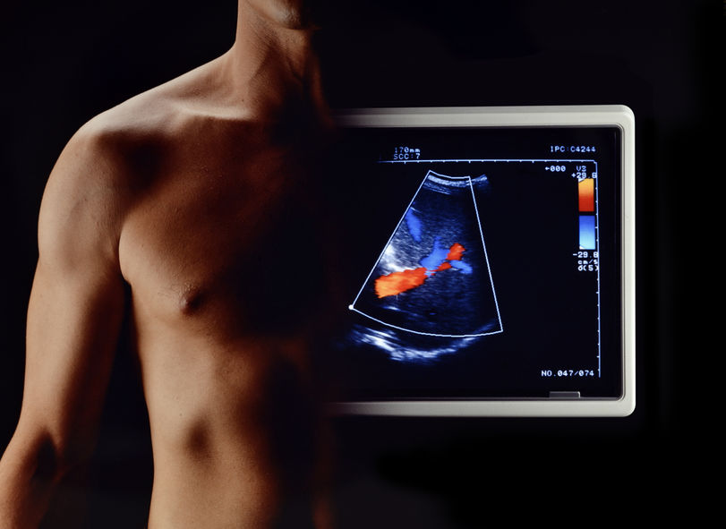
An echocardiogram (often called "echo") is a graphic outline of the heart's movement. During this test, high-frequency sound waves, called ultrasound, provide pictures of the heart's valves and chambers. This allows the echo technician to evaluate the pumping action of the heart. Echo is often combined with Doppler ultrasound and color Doppler to evaluate blood flow across the heart's valves. The echocardiogram procedure uses the same technology used to evaluate a baby's health before birth. A hand-held device called a transducer is placed on the chest and transmits high frequency sound waves (ultrasound). These sound waves bounce off the heart structures, producing images and sounds that can be used by the doctor to detect heart damage and disease..
Echocardiography is an invaluable tool in providing the doctor with important information including size of the chambers of the heart, pumping function of the heart, and structure and function of each heart valve.
Stress Echo-Cardiogram
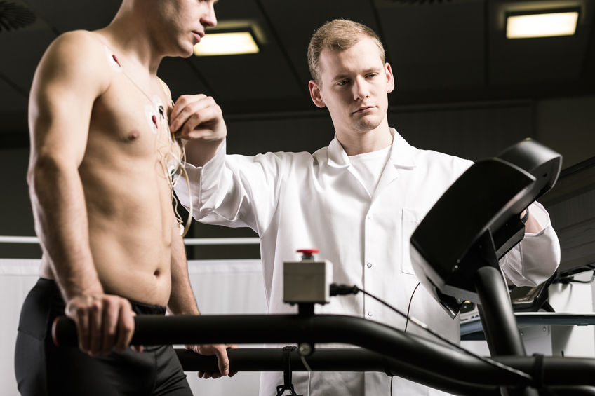
This is an echocardiogram that is performed in combination with exercise on a treadmill. This test can be used to visualize the motion of the heart's walls and pumping action when the heart is stressed. It may reveal a lack of blood flow that isn't always apparent on other heart tests.Exercise stress testing usually employs the "Bruce" protocol, Exercise is started at a slower "warm-up" speed. The speed of the treadmill and it's slope or inclination is increased every 3 minutes. The treadmill is usually stopped when the patient exceeds 85% of the target rate (based upon the patient's age).
EKG recordings are made during every minute of exercise and then again after exercise is stopped. Immediately after stopping the treadmill, the patient moves directly to the examination table and lays on the left side. The Echo examination is immediately repeated. A video clip of multiple views of the resting and exercise study are compared side-by-side and analyzed by the physician.
Holter monitoring And Arrythmia Detection
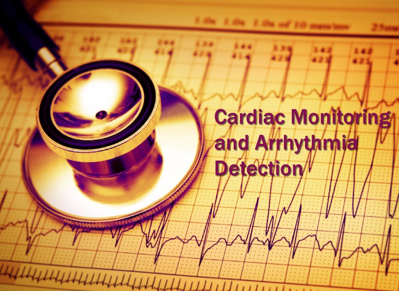
A Holter monitor is a small, wearable device that you usually wear for 24-hours, and during that time, the device will record all of your heartbeats. The Holter monitor has electrodes that are attached to your chest with adhesive and then are connected to a recording device. The doctor uses information captured on the Holter monitor's recording device to figure out if you have a heart rhythm problem.
Ambulatory Blood Pressure Monitoring
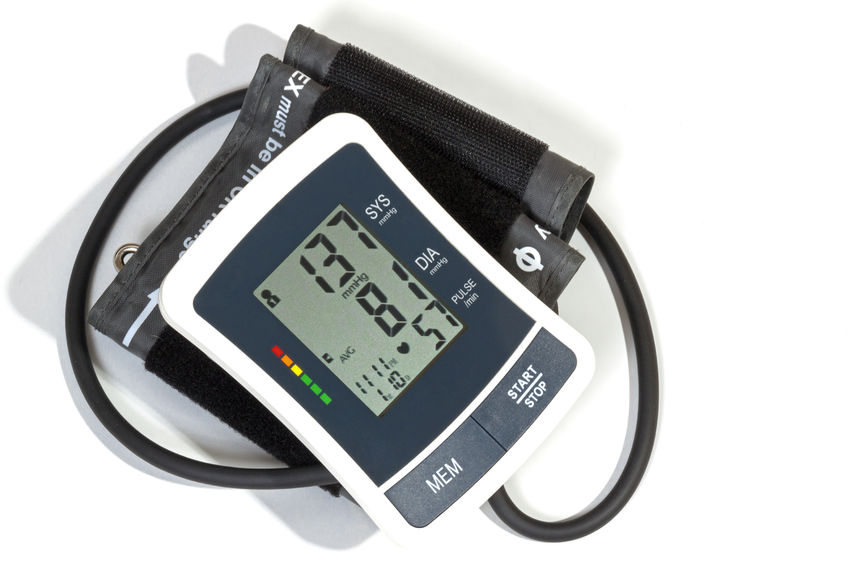
Ambulatory Blood Pressure Monitoring An ambulatory blood pressure monitor is a small device which you wear on a belt. The blood pressure cuff, worn under your clothes, is connected to the device and automatically checks your blood pressure every 1-2 hours, even while you are sleeping. After 24 hours of monitoring, you will take the device to the doctor's office. The blood pressure information is transferred from the device to a computer. Your doctor will review the information with you and decide if your treatment program is working or if you need to make changes.
Other Services Provided In Hospital
Dr. Agarwal is Level-II Certified in Coronary CT-Angiography and can provide this service at the Medical Center of Plano when deemed clinically appropriate.

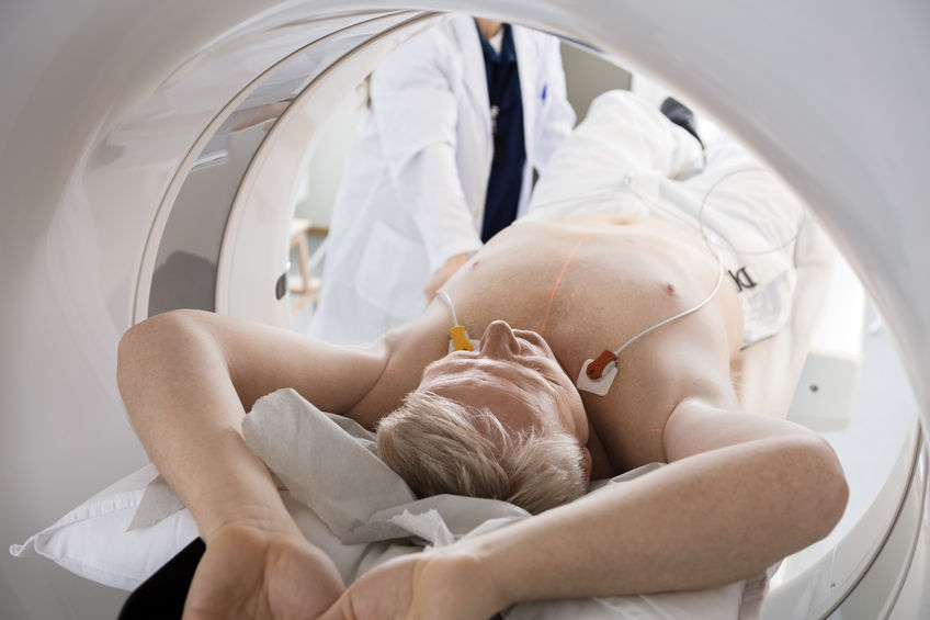
A coronary computed tomography angiogram uses advanced CT technology, along with intravenous (IV) contrast material (dye), to obtain high-resolution, three-dimensional pictures of the moving heart and great vessels. CTA is used as a noninvasive method for detecting blockages in the coronary arteries.
Dr. Agarwal also performs diagnostic Cardiac Catheterization at the Medical Center of Plano hospital and Baylor Heart Hospital to determine the extent of coronary artery disease for possible coronary intervention or bypass surgery.
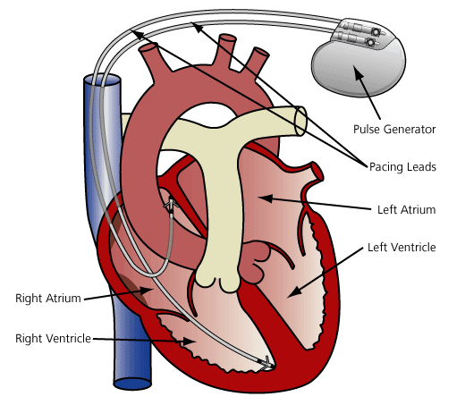
Other procedures performed by her at the hospital include Pacemaker implantation, Trans-Esophageal Echocardiogram, and Cardioversion.
The Pacemaker is a battery-powered implantable devices that uses electrical pulses to prompt the heart to beat at a normal rate. Pacemakers are used to treat arrhythmias (ah-RITH-me-ahs). Arrhythmias are problems with the rate or rhythm of the heartbeat. During an arrhythmia, the heart can beat too fast, too slow, or with an irregular rhythm.

